Alicja E Kownacka1, Benjamin P Burke1, Juozas Domarkas1, Mark Lorch1, Martin Pickles1, Peter Gibbs1, Joan Simo2,3, Chris Wilson2, Alan Rowan3, Simone Mastrogiacomo3, Frank Walboomers3 and Stephen J Archibald1
1University of Hull, UK 2NovioTech BV, The Netherlands 3University of Nijmegen, The Netherlands
Scientific Tracks Abstracts: J Bioengineer & Biomedical Sci
Visualisation of tissue engineering constructs in vivo is required in order to monitor their behaviour and integration into surrounding tissues or to promptly detect immune rejection. Various analytical tools could be used to that end, however, most of them require invasive biopsies and allow only a single time point characterisation. Magnetic resonance imaging (MRI) is a non-invasive technique, therefore, its potential usage in tissue engineering area is a major focus of research effort. Nevertheless, MRI suffers from poor sensitivity, thus, there is a clear need for the development of novel contrast agents and also for elaboration of methodologies allowing their conjugation to the engineered materials. Presently, biomedical hydrogels used for soft tissue reconstructions cannot be distinguished from a normal tissue by MRI due to their high water content. Similarly, calcium phosphate cements (CPCs) designed for bone regeneration are too dense and do not contain the required amount of water for MR imaging. These limitations can be overcome by use of stable metal chelates or nanomaterials as contrast agents. Current work on gadolinium complexes functionalised for chemospecific conjugation with polyisocyanopeptide hydrogels and development of bone targeted gadolinium nanoparticles will be presented.
Alicja E Kownacka was awarded MSc in Biomaterials degree from Westpomeranian University of Technology, Poland, where she worked on photo curable polymeric systems for novel hernia treatment. For the following year, she worked in 3 different European Institutes developing nanomaterials for various applications including MR imaging. In 2014, she won a Marie-Curie PhD scholarship and joined Prof. Steve Archibald’s group at University of Hull, UK, where she is currently working on a non-invasive visualisation of soft and hard tissue engineering constructs using Magnetic Resonance Imaging (MRI) and Positron Emission Tomography (PET).
Email: A.Kownacka@hull.ac.uk
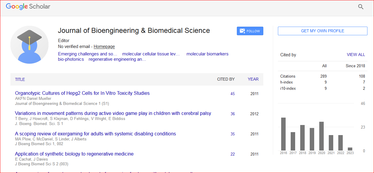
Journal of Bioengineering & Biomedical Science received 307 citations as per Google Scholar report


 Spanish
  Chinese
  Russian
  German
  French
  Japanese
  Portuguese
  Hindi
Hindi 