Hideo Akiyoshi, Asuka Inoue-Matsuo and Itaru Onodera
This study presented detailed descriptions of parenchymal arrangements in the livers of 23 reptilian species using light microscopy, and extensively discussed this from a phylogenetic viewpoint. Hepatocyte sinusoidal structures (HSS) were classified into three different types: (I) the several-cell-thick plate type, (II) two-cell-thick plate type, and (III) one-cell-thick plate type. Parenchymal arrangements showed either the combined two- and one-cell-thick plate type or one-cell-thick plate type, whereas the sea snake in the sub-order Serpentes showed the several-cell-thick plate type. In the order Testudines, peripheral sinusoids near terminal portal veins were tortuous, becoming straighter toward terminal central veins. Melanomacrophages (MMs) were observed in sinusoidal capillaries in the order Testudinata, Crocodilia, and Squamata, but not in the sub-order Serpentes (except for the sea snake). This study showed that the architecture of the parenchymal arrangement was related to the phylogenetic relationship, whereas the distribution of MMs may not be. The MMs systems of turtles, alligators, and sea snakes, whose place of main habitation is underwater, may have adapted according to ecological and behavioral patterns. Based on the hepatic architecture of parenchymal arrangements and the distribution of MMs, it was suggested that reptilian livers acquired the division of three zones in the acinus.
PDFShare this article
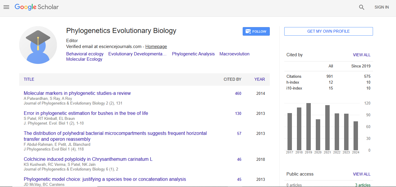
Journal of Phylogenetics & Evolutionary Biology received 911 citations as per Google Scholar report