|
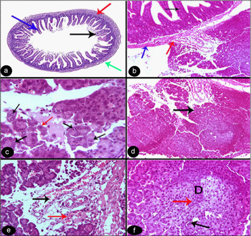
| Figure 1: (a) a photomicrograph of group (1) showing normal histological structure of the jejunum, intestinal villi (black arrow), lamina propria (blue arrow), crypt of Lieberkühn (red arrow) and tunica muscularis (green arrow). Stain: H&E. Mag. X 4. (b): a photomicrograph of a section in the intestine group (2) showing intestinal villi (black arrow), tunica muscularis (red arrow) and blood vessels (blue arrow). Stain: H&E. Mag. X 10. (c): a photomicrograph of group (2) showing cells with hyperchromatic nuclei with typical (red arrow) and atypical (black arrow) mitotic figures. Stain: H&E. Mag. X 40. (d): a photomicrograph of group (2) showing cells with different shape and size, mostly ovoid to polyhedral in shape (arrow). Stain: H&E. Mag. X 10. (e): a photomicrograph of group (2) showing congested blood vessels (black arrow) and focal hemorrhage in the stroma (red arrow). Stain: H&E. Mag. X 25. (f): a photomicrograph of group (2) showing the center of the neoplastic mass with some degenerated and necrotic cells (D), some cells with pycknotic (black arrow) or karryolytic (red arrow) nuclei. Stain: H&E. Mag. X 25. |