| Research Article |
Open Access |
|
| Asha KR*, Vinay Kumar K and Lakshmi Prabha Subhash |
| Department of Anatomy, Sri Siddhartha Medical College, Tumkur, Karnataka, India |
| *Corresponding author: |
Dr. Asha KR
Associate Professor
Department of Anatomy
Sri Siddhartha Medical College
Tumkur, Karnataka, India
E-mail: ashakeshavraj@rediffmail.com |
|
| Â |
| Received September 15, 2012; Published December 03, 2012 |
| Â |
| Citation: Asha KR, Vinay Kumar K, Subhash LP (2012) Analysis of Anthropometric Indices in Down Syndrome Children. 1:522. doi:10.4172/scientificreports.522 |
| Â |
| Copyright: © 2012 Asha KR, et al. This is an open-access article distributed under the terms of the Creative Commons Attribution License, which permits unrestricted use, distribution, and reproduction in any medium, provided the original author and source are credited. |
| Â |
| Abstract |
| Â |
| Objective: To determine in a representative sample of young patients with Down syndrome, the specificity of anthropometric profile for the syndrome, which can be used in the diagnosis, monitoring of growth and to identify anthropometric variables which best discriminate group of patients with Down syndrome from healthy persons. Limited Asian reports are available, and may not be able to be extrapolated for use in our local population, due to a differing mix of ethnicities. Hence the present study was conducted to analyze the anthropometric characteristics of Down syndrome in South Indian patients. |
| Â |
| Methods: The present study was conducted on 100 subjects of South Indian origin. Using non-invasive method of anthropometry, fifteen anthropometric measurements were performed and eleven indices were calculated in 50 Down syndrome patients and 50 age and sex matched controls, aged 1-18 years. |
| Â |
| Results: Stepwise forward discriminant function analysis identified a subset of five variables namely body mass index, cephalic index, index of size of head, morphological upper facial index and foot index which could accurately classify subjects with Down syndrome. |
| Â |
| Conclusion: Anthropometric analysis of Down syndrome children revealed their specific characteristics since they reflect the influence of environmental factors and genetic determinants. Results from the present study show that trisomy 21 has a clearly defined and different phenotype than the healthy population as early as childhood. |
| Â |
| Keywords |
| Â |
| Down syndrome; Anthropometry; Trisomy 21 |
| Â |
| Introduction |
| Â |
| Down syndrome (Trisomy 21) is one of the commonest chromosomal disorders which occur in one in 650-1000 live births. Mental retardation, dysmorphic facial features and other distinctive phenotypic traits characterize the Down syndrome [1]. A precise diagnosis makes available all the accumulated knowledge and experience of that condition and generally provides a better estimate of the risk of recurrence. It informs prognosis and permits interventions that may prevent, anticipate or more successfully treat complications. Furthermore, an accurate diagnosis is the key to research into the identification of causative genes, interventions and treatments [2]. Diagnostic accuracy would be improved by objective quantitative criteria and analytic methodology where possible [3]. |
| Â |
| Most syndrome diagnoses are suggested by the gestalt of the patient, in the same way that most people would recognize a child with Down syndrome. Notwithstanding an initial impression, it is necessary to perform a detailed examination and compare the observations and history with those expected in the syndrome being considered or with the lead suggested by the database. It is important not to rush too early to a diagnosis because, once applied to a patient's condition, labels are hard to remove. Concerns about the subjectivity of some syndrome diagnoses, especially at the mild end of a syndrome's spectrum, have led some dysmorphologists to explore more objective diagnostic approach such as anthropometrics from standardized physical landmarks, to assess patients objectively [2]. |
| Â |
| Anthropometry has much to offer the clinical geneticist because it is simple and non-invasive, with minimal equipment [3]. There is a widespread agreement that anthropometry is the technique of choice for the evaluation of dysmorphic features. It is estimated that about 95% of patients who are suspected of Down syndrome can be categorized with 99.9% confidence by anthropometric measurements. One can thereby make a fast clinical diagnosis on the majority of suspects before karyotyping is complete [1]. |
| Â |
| The purpose of the present study was aimed at studying the various anthropometric measurements in patients of Down syndrome, so that the characteristic clinical features can be studied specifically by comparing with the age and sex matched control group. The study also aims to establish the anthropometric variables which discriminate Down syndrome from healthy population. |
| Â |
| Materials and Methods |
| Â |
| Fifty South Indian Down syndrome children (30 males and 20 females) aged 1-18 years were chosen as a defined population to investigate application of anthropometric craniofacial pattern profiles. Control group consisted of fifty healthy individuals of the same age, ethnicity and gender. The evaluation of measurements from children with Down syndrome and normal children from the same ethnic background eliminated the population specific variations. All the controls included in the study were healthy with adequate nutrition and did not have any congenital malformations. |
| Â |
| In a pretested performa, detailed history and general physical examination of the subjects including anthropometric measurements meeting the objectives of the study were taken. The subjects were examined after informed written consent from their parents or guardians was obtained. |
| Â |
| A total of 15 anthropometric measurements per subject were performed by single observer in order to avoid inter-observer bias. Measurements were recorded following the methodology according to Singh [4] using anthropometer, weighing scale, sliding caliper, spreading caliper, first segment of anthropometer rod and flexible inelastic tape. |
| Â |
| Anthropometric measurements |
| Â |
| Stature: It was measured as vertical distance from vertex (v) to floor using anthropometer; |
| Â |
| Weight: measured using weighing scale. |
| Â |
| Measurements on the head |
| Â |
| |
| Maximum head length: measured as straight distance between glabella (g) and opisthocranion (op) using spreading caliper; |
| Â |
| Maximum head breadth: measured as straight distance between the two eurya (eu) using spreading caliper; |
| Â |
| Breadth of bizygomatic arch: measured as distance between two zygia (zy) i.e., the most lateral points on the zygomatic arch using spreading caliper; |
| Â |
| Head height: measured as projective distance between apex (apx) and tragion (t) using first segment of anthropometer rod or rod compass with head height needle. |
| Â |
| Measurements on the face |
| Â |
| Morphological facial height: measured as straight distance between nasion (n) and gnathion (gn) using sliding caliper; |
| Â |
| Morphological upper facial height: measured as straight distance between nasion (n) and prosthion (pr) using sliding caliper; |
| Â |
| Physiognomic ear length: measured as straight distance between superaurale (sa) and subaurale (sba) i.e., highest and lowest points on the external ear using sliding caliper. |
| Â |
| Physiognomic ear breadth: measured as straight distance the most lateral points i.e., preaurale (pra) and postaurale (pa) when taken at right angles to the physiognomic ear length using sliding caliper. |
| Â |
| Measurements on the hand |
| Â |
| Length of hand: measured as straight distance between the midpoint of a line joining the two stylion (sty) and dactylion (da) of the middle finger using sliding caliper; |
| Â |
| Breadth of hand: measured as straight distance between metacarpal radiale (mr) and metacarpal ulnare (mu) using sliding caliper. |
| Â |
| Measurements on the foot |
| Â |
| Length of foot: measured as straight distance directly from pterion (pte) to acropodion (ap) using first segment of anthropometer rod; |
| Â |
| Breadth of foot: measured as straight distance between metatarsal tibiale (mtt) and metatarsal fibulare (mtf) using sliding caliper. |
| Â |
| Measurement on the chest |
| Â |
| Chest circumference: measured as horizontal circumference of the upper part of the body trunk at the level of mesosternal in the resting stage using flexible inelastic tape. |
| Â |
| From the above measurements following indices were calculated |
| Â |
1. |
| Â |
2.  |
| Â |
| 3. Index of size of head: Maximum head length × Maximum head breadth× Head height |
| Â |
4.  |
| Â |
5.  |
| Â |
6.  |
| Â |
7.  |
| Â |
8.  |
| Â |
9.  |
| Â |
10.  |
| Â |
11.  |
| Â |
| Statistical Results |
| Â |
| The data was analyzed using the software SPSS (Statistical Package of Software System) version 12.0. |
| Â |
| The indices calculated from various anthropometric measurements were subjected to the following Statistical methods (Table 1). |
| Â |
|
|
Table 1: Independent samples‘t’ test of anthropometric indices in Down syndrome patients and controls. |
|
| Â |
| • Independent samples t test. |
| Â |
| • Discriminative analysis. |
| Â |
| Discriminative Analysis |
| Â |
| Stepwise Discriminant Function Analysis was adopted to study variables which contribute independently in Discriminant Analysis. Probability at 0.05 levels was used for studying contribution of each of the independent variables. |
| Â |
| ‘F’ test is used at each level of introducing new variable for Discriminant Function. The Eigen value of each Discriminative function analysis reflects the ratio of importance of the dimensions which classify cases of the dependent variables (Tables 2 and 3). |
| Â |
|
|
Table 2: Showing the Eigen Value. |
|
| Â |
|
|
Table 3: Showing Wilk’s Lambda. |
|
| Â |
| Unstandardized discriminant coefficients were used in the formula for making the classifications in Discriminative function analysis (Table 4). |
| Â |
|
|
Table 4: Canonical Discriminant Function Coefficient. |
|
| Â |
| In the present study, total of 11 variables were used initially. Stepwise Discriminant Function Analysis could include only 5 variables namely Cephalic index, Index of size of Head, Morphological upper facial index, Body mass index and Foot index which were independently contributing to Discriminant Function Analysis. |
| Â |
| The formula for classifying the Down syndrome from control group was as follows: |
| Â |
| -11 + (Cephalic index × 0.049) + (Index of size of Head × 0.001) + (Morphological upper facial index × -0.076) + (Body Mass Index × 0.127) + (Foot Index × 0.150). If obtained value from the above formula is less than 3.151, then it belongs to group 1 i.e., Down syndrome patients. If it is greater than 3.151, it belongs to group 2 i.e., controls. |
| Â |
| To measure the strength of relationships, classification table was used. It assesses the performance of Discriminative function analysis (Table 5). |
| Â |
|
|
Table 5: Showing classification results. |
|
| Â |
| Discussion |
| Â |
| Subjects with DS provide a useful model for investigating the effect that chromosomal aneuploidy has on normal development. The deviant physical development in DS can be effectively illustrated anthropometrically. Clinical findings corroborate the theory that the specificities found in persons with DS are the result of irregular development and growth in the early embryonic period [5]. This represents persistence of infantile body proportions [6]. |
| Â |
| The results in the present study depict higher values for males in all parameters measured. These can be explained on the basis the physical personality of the individual has a bearing on the anthropometric landmarks (Supplementary file) so the male characteristics are usually larger [7]. Diagnosis of congenital anomaly like DS usually involves clinical observation of morphological findings and abnormal body proportions [1]. |
| Â |
| The present study was aimed to establish the various anthropometric variables that discriminate 50 patients of Down syndrome from 50 age and sex matched controls. From 15 anthropometric measurements, 11 indices were calculated as mentioned earlier. |
| Â |
| Stature |
| Â |
| Styles et al. states that short stature is a recognized characteristic of most people with DS. Average height at most ages are around 2nd centile for the general population [8]. |
| Â |
| Cronk et al. studied growth rate of DS children for 2 age intervals, 1-36 months and 2-18 years. They provide the data which corroborate other studies of growth in children with DS demonstrating deficient growth rate throughout the growing period [9]. |
| Â |
| Farkas et al. performed cross-sectional study in 3 age categories in 115 DS patients in 1-36 years old to prove short stature as stigmata of DS [10]. |
| Â |
| In the present study, when height of the subjects were referred to standard pediatric growth charts, it revealed that height in most of the DS children was under 3rd to 10th percentile and controls under 25th to 75th percentile. Our findings are consistent with the findings of earlier studies. |
| Â |
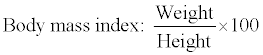
| Body mass index |
| Â |
| Cronk et al. found that DS children have a tendency to be overweight beginning in late infancy and throughout the remainder of the growing years [9]. |
| Â |
| Myrelid et al. showed that in DS children, Body mass index more than 25 Kg/m2 at 18 years of age was observed in 31% of the males and 36% of the females [11,12]. |
| Â |
| The present study in which BMI of Down syndrome patients was nearer to upper limit of normal range differed from the above mentioned studies. However our study correlated with that of Styles et al. [8] which states that people with the Down syndrome are not necessarily overweight in relation to their height (Table 6). |
| Â |
|
|
Table 6: Classification of children according to BMI [13]. |
|
| Â |
| Regular growth surveillance of children with Down syndrome should aid early identification both of pathological causes of growth retardation and incipient overweight or obesity. Growth charts are recognized as a useful tool for monitoring the growth and well being of children [8]. |
| Â |
| Cephalic index |
| Â |
| Brachycephaly is included in Jackson list of 25 signs of DS [13]. The present study on Cephalic index correlated with study of Allanson et al. who did a series of 21 anthropometric craniofacial measurements [14]. |
| Â |
| In the present study, increase in Cephalic index in DS group (88) was found to be significantly increased than in controls (76). |
| Â |
| Accordingly, DS patients were classified under brachycephalic group (cephalic index-88) and controls under mesocephalic group (cephalic index-76) (Table 7). |
| Â |
|
|
Table 7: Range variation of Cephalic index according to Singh [4]. |
|
| Â |
| Index of size of head |
| Â |
| Review of literature indicates that DS patients are characterized by microcephaly [14,15]. In the present study the mean value of Index of size of the head in DS patients (830) was decreased than control group (899). |
| Â |
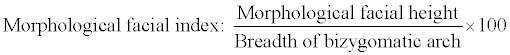
| Morphological facial index |
| Â |
| The study by Farkas et al. showed that highest percentage of mild to moderate disproportion is found in the face [16]. |
| Â |
| The study by Sforza et al. showed that independently of sex, subjects with DS had faces that were significantly narrower, less deep and shorter than the faces of normal subjects [17]. |
| Â |
| The present study correlated with the above mentioned studies by revealing a statistically significant decrease in the Morphological facial index in DS patients (75) compared to controls (87) (Table 8). |
| Â |
|
|
Table 8: Range variation of Morphological facial index according to Singh [4]. |
|
| Â |
| Accordingly, DS patients could be classified under Hypereuryprosopic group and controls under Mesoprosopic group. |
| Â |
| Morphological upper facial index |
| Â |
| The study by Allanson et al. confirmed that decreased upper facial depth was one of the variables which could accurately classify more than 99% of the individuals in the combined sample of affected and unaffected individuals [14]. |
| Â |
| The study by Farkas et al. proved that the highest percentage of mild-moderate disproportion was found in the face (79.3%) and found that a shallow upper third of the face depth was the most frequent subnormal finding (71.5%) [18]. |
| Â |
| In the present study, mean value of Morphological upper facial index in DS patients was 37 and in controls it was 52. This decrease in the Morphological upper facial index in DS patients was statistically significant when compared with controls (Table 9). |
| Â |
|
|
Table 9: Range variation of morphological upper facial index according to Singh [4]. |
|
| Â |
| Accordingly, DS patients could be classified under Hypereuren group and controls under Mesen group. |
| Â |
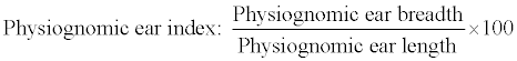
| Ear index |
| Â |
| The study by Allanson et al. confirmed decreased ear length as one of the profiles which could accurately classify more than 99% of individuals in the combined sample of affected and unaffected individuals [14]. |
| Â |
| In the study by Farkas et al. short right auricle was the most frequent subnormal finding in 52 DS patients (71.5%) [18]. |
| Â |
| The study by Chou et al. concluded that decreased ear length is important in diagnosing DS patients. They studied 20 patients of DS and found that ear length of patients of DS was indeed smaller than that of normal children [3]. |
| Â |
| The study by Sforza et al. revealed that subjects with DS had larger ear width-to-ear length ratios [17]. |
| Â |
| The study by Ferrario et al. showed that in DS patients’ ear width and ear length were significantly reduced [19]. |
| Â |
| The present study correlated with the study by Sforza et al. [17] and Chou et al. [3] revealing that Ear index in DS patients (78) was significantly increased than controls (73). |
| Â |
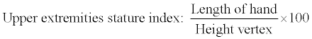
| Upper extremity stature index |
| Â |
| Shortness of upper extremities is typical of DS patients [20]. In the present study, upper extremity stature index was decreased in DS patients (11.15) than in controls (11.23) though statistically insignificant. |
| Â |
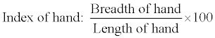
| Index of hand |
| Â |
| Hand in DS patients is short and broad i.e., handbreadth is more than hand length [16,20]. In the present study, Index of hand was significantly increased in DS patients than controls. |
| Â |
| In DS patients mean value of Index of hand was 54 and in controls 44 (Table 10). |
| Â |
|
|
Table 10: Range variation of Hand index. |
|
| Â |
| Accordingly, DS patients were classified under Hyperbrachycheir group and controls under Mesocheir group. |
| Â |
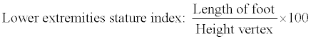
| Lower extremity stature index |
| Â |
| In DS patients, shortness of lower extremities is one of the features [20]. In the present study, lower extremity stature index was decreased in DS patients (14.6) than in controls (14.8). But, this difference is statistically insignificant. |
| Â |
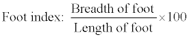
| Foot index |
| Â |
| DS patients are characterized by broad and short foot [20]. In the present study, mean value of Foot index (48) in DS patients was significantly more than that of control group (40) (Table 11). |
| Â |
|
|
Table 11: Range variation of Foot index. |
|
| Â |
| Accordingly, DS could be classified under Brachypod group and controls under Mesopod group. |
| Â |
| Chest circumference index |
| Â |
| This index was not included in any of the previous studies. In the present study, a significant increase in Chest circumference of DS patients was observed. Mean value of CCI in DS patients was 58 and in controls, 51 (Table 12). |
| Â |
|
|
Table 12: Range variation of Chest circumference index. |
|
| Â |
| Accordingly, DS patients could be classified as having broad chest and controls with medium shoulders. |
| Â |
| Anthropometric study by Allanson et al. [14] revealed that Stepwise forward discriminant function analysis identified a subset of 3 variables (ear length, maxillary arch and upper facial depth) that could accurately classify more than 99% of the individuals in the combined sample of affected and unaffected individuals. Of the subjects with Down syndrome, 96.8% were classified correctly. |
| Â |
| Study by Bagic and Verzak [5] showed that three variables head length, head circumference and outer canthal distance were responsible for 85.68% variability (p<0.001). |
| Â |
| In our study, Stepwise Discriminant Function Analysis could include 5 variables namely Cephalic index, Index of size of head, Morphological upper facial index, Body mass index and Foot index which were independently contributing to Discriminant Function Analysis. |
| Â |
| Within the limitation of methodology and sample size differences, most variables except breadth of head, ear, hand and foot were smaller in subjects with DS than their normal controls selected for sex, age and ethnicity. This study indicates the possibility of applying anthropometric measurement of variables for producing an anthropometric profile, which gives a completely objective picture of that which is determined by clinical examination and inspection. |
| Â |
| Conclusion |
| Â |
| On the basis of anthropometric measurements of South Indian origin, it was possible to conclude that persons with DS have a specific and recognizable anthropometric pattern with specifically expressed deviations from normal controls of the same age, sex and ethnic group, which enable their discrimination from healthy persons. The indices allowed discriminating 94% of subjects with DS when compared with normal subjects. Obviously, there is a need for a larger, more representative sample to resolve some differences in a variety of ethnic and age structure classification interpretations encountered in the literature. |
| Â |
| |
| References |
| Â |
- Rex AP, Preus M (1982) A diagnostic index for Down syndrome. J Pediatr 100: 903-906.
- Hunter AG (2002) The diagnostic approach to the child with dysmorphic signs. CMAJ 167: 367-372.
- Chou CT, Tseng YC, Tsai FJ, Lin CC, Liu CS, et al (2002) Measurement of ear length in neonates, infants and preschool children in Taiwan. Acta Pediatr Taiwan 43: 40-42.
- Singh IP (2004) Anthropometry: A Laboratory Manual on Biological Anthropology. (1stedn), Kamla Raj Enterprises, India.
- Bagic I, Verzak Z (2003) Craniofacial anthropometric analysis in Down's syndrome patients. Coll Antropol 2: 23-30.
- Jaswal S, Jaswal IJ (1981) An anthropometric study of body size in Down syndrome. Indian J Pediatr 48: 81-84.
- Agnihotri G, Singh D (2007) Craniofacial anthropometry in Newborns and Infants. Iran J Pediatr 17: 332-338.
- Styles ME, Cole TJ, Dennis J, Preece MA (2002) New cross sectional stature, weight, and head circumference references for Down's syndrome in the UK and Republic of Ireland. Arch Dis Child 87: 104-108
- Cronk C, Crocker AC, Pueschel SM, Shea AM, Zackai E, et al (1998) Growth charts for children with Down syndrome: 1 month to 18 years of age. Pediatrics 81: 102-110.
- Farkas LG, Katic MJ, Forrest CR (2002) Age-related changes in anthropometric measurements in the craniofacial regions and in height in Down's syndrome. J Craniofac Surg 13: 614-622.
- Myrelid A, Gustafsson J, Ollars B, Annerén G (2002) Growth charts for Down's syndrome from birth to 18 years of age. Arch Dis Child 87: 97-103.
- Park K (2005) Epidemiology of chronic non-communicable diseases and conditions. Text book of Preventive and Social Medicine. (18thedn), Banarsidas Bhanot Publishers, India.
- Avramopoulos D, Kennerknecht I, Barbi G, Eckert D, Delabar JM, et al. (1997) A case of apparent trisomy 21 without the DownÂ’s syndrome phenotype. J Med Genet 34: 597-600.
- Allanson JE, O'Hara P, Farkas LG, Nair RC (1993) Anthropometric craniofacial pattern profiles in Down syndrome. Am J Med Genet 47: 748-752
- Ghai OP, Piyush Guptha, Paul VK (2004) Genetic disorders. Ghai Essential Pediatrics. (6thedn), CBS Publishers and distributors, India.
- Farkas LG, Katic MJ, Forrest CR (2001) Surface anatomy of the face in Down's syndrome: anthropometric proportion indices in the craniofacial regions. J Craniofac Surg 12: 519-524.
- Sforza C, Dellavia C, Tartaglia GM, Ferrario VF (2005) Morphometry of the ear in DownÂ’s syndrome subjects. A three-dimensional computerized assessment. Int J Oral Maxillofac Surg 34: 480-486.
- Farkas LG, Munro IR, Kolar JC (1985) Abnormal measurements and disproportions in the face of Down's syndrome patients: preliminary report of an anthropometric study. Plast Reconstr Surg 75: 159-169.
- Ferrario VF, Dellavia C, Zanotti G, Sforza C (2004) Soft tissue facial anthropometry in Down syndrome subjects. J Craniofac Surg 15: 528-532.
- Kulkarni ML (2006) Genetic disorders, IAP Textbook of Pediatrics. (3rdedn), Jaypee Brothers Medical Publishers, India.
|
| Â |
| Â |