 |
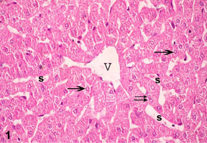
| Figure 1: Photomicrographs of sections in the liver of control group I showing anastomosing cords of hepatocytes radiating from central vein (V) separated by blood sinusoids (s) and the hepatocytes contains central pale stained nuclei (arrows) and some are binucleated (double arrow). |