 |
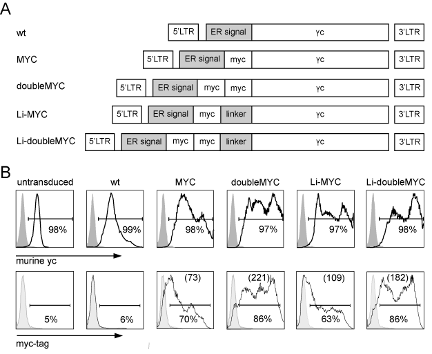
| Figure 1: Variants of the myc-tagged murine γc are expressed on 58 cells and are differentially detected by anti-myc ab. (A) Composition of retroviral vectors encoding wt murine γc and differently tagged variants. The DNA sequences for the murine γc chain or its variants with single or double myc-tag and with or without linker were inserted into the retroviral vector MP71. (B) 58 cells were transduced with wt or myc-tagged murine γc variants shown in (A). γc surface expression was quantified by flow cytometry using antimurine γc (upper panel) and anti-myc ab (lower panel). Tinted curves show isotype control staining (for anti-γc ab) or staining of untransduced cells (for myc-specific ab), respectively. Numbers in brackets indicate the MFI of mycspecific staining in the myc-positive population. (LTR: long terminal repeat, ER: endoplasmatic reticulum). |