 |
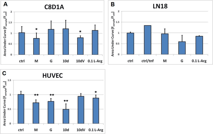
| Figure 2: Effects of exogenous humanins on the intracellular calcium release. The cells were incubated with selected HNM, HNG, HN10d, HN10dV for 24 hours, followed by either stimulation with 25 μM βA for one minute (brain cells) or 5 ng/mL TNFα (endothelial cells). The calcium release was induced by 10 mM ATP, and changes in Fura2 fluorescent ratios were recorded for 40 seconds. The results were calculated as average area under curve derived from continuous recording of Fura2 ratios. The results were normalized to the signal generated by the control cells; the experiments were done in triplicates at minimum. A) C8D1A cells, B) LN18 cells, C) HUVECs. |