 |
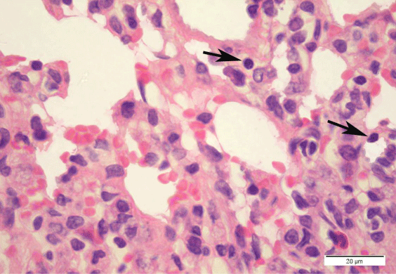
| Figure 17: A photomicrograph of a DEHP recovery rat alveolar tissue showing extravasated blood cells in alveolar lumens, with less inflammatory cell infiltration. Some necrotic type II pneumocytes with pyknotic nuclei are shown (black arrow). (H & E x1000). |