 |
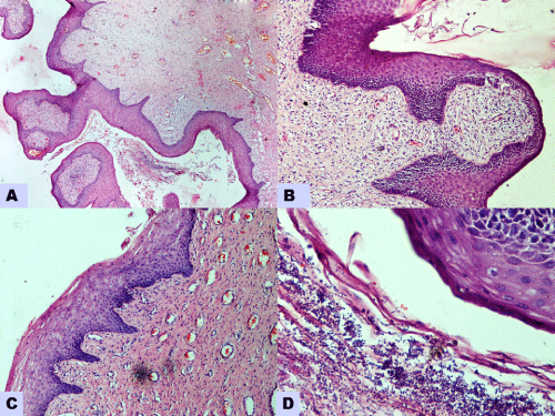
| Figure 2: Microscopic examination showing (A): Polyp covered by stratified squamous epithelium and underlying vascularized stroma (H& E stain, 40x), (B): Stromal chronic inflammatory infiltrate (H& E stain, 100x), (C):Koilocytic changes of stratified squamous epithelium (H& E stain, 100x) and (D): Yeasts and pseudohyphae of fungus (H& E stain, 400x). |