 |
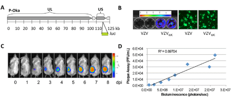
| Figure 2: Construction of recombinant VZV containing a luciferase gene (VZVLUC) A. To generate a VZVLUC strain, a luciferase expression cassette was inserted into the intergenic region between ORF65 and ORF66 of the VZVLUC genome. This clone was then transfected into MeWo cells to produce the VZVLUC strain. B. MeWo cells were grown in six-well culture dishes and infected with VZV BAC or VZVLUC BAC. Bioluminescence from VZVLUC infected cells was then visualized and measured using IVIS (Xenogen) imaging. Strong bioluminescence was detected only from VZVLUC -infected cells. In addition, green fluorescent plaques were observed for all wells using fluorescence microscopy, indicating that both viruses were infectious. C. Example of in vivo IVIS imaging for growth curve analysis in a mouse VZV model. Image adapted from Zhang et al. [10]. D. Correlation between traditional viral plaque assay and luciferase-based IVIS bioluminescence growth analyses for accurate estimation of viral titer. |