 |
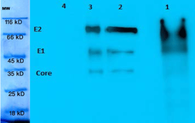
| Figure 12: Western blot of recombinant Core (20 kDa), E1(40 kDa), E2 (60 kDa). (1) Recombinant protein before PNGaseF and it is smear like and heavy with N-glucan bands. (2) Recombinant protein after PNGaseF without extra N-glucan linkages and sharp bands. (3) HCV proteins as Positive control. (4) Negative control. MW is molecular weight markers. |