 |
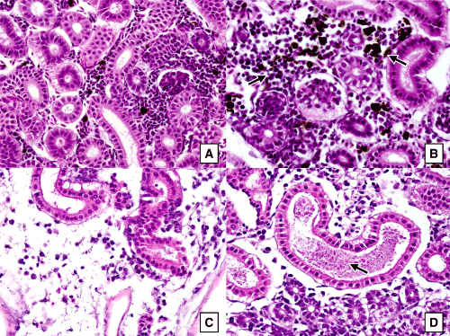
| Figure 3: Sections of kidney tissues of coho salmon fry (three months old) stained with H&E and magnified 400X showing: (A) healthy kidney tissues of coho salmon from the negative control group, (B) kidney tissues of an intraperitoneally infected fish with melanomacrophage hyperplasia, (C) kidney tissues of an intraperitoneally infected fish with tubular degenerative changes and edema within the renal interstituim, (D) kidney tissues of an intraperitoneally infected fish with renal tubular degeneration and proteinaceous casts in the tubular lumen (arrow) |