 |
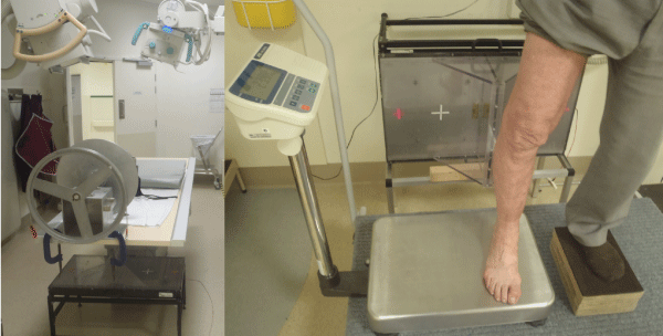
| Figure 3: (a) Torsional DLRSA setup with radiographic tubes angled at 30 degrees from perpendicular to the calibration cage. (b) Axial loading DLRSA setup with the weightbearing foot positioned on the scales and the RSA calibration cage behind the patient. |