 |
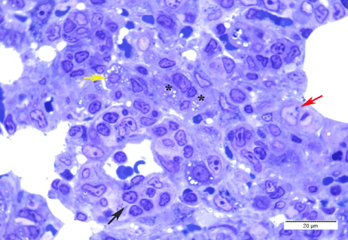
| Figure 12: A photomicrograph of a semithin section of a DEHP treated rat alveolar tissue showing loss of normal alveolar architecture in thickened interalveolar septum, increased number of alveolar macrophages surrounding collapsed alveoli (black arrow). Large numbers of type II pneumocyte are shown, some with chromatin margination (yellow arrow); karyolytic nuclei (*) and enlarged pale nucleus (red arrow). (Toluidine blue x1000). |