 |
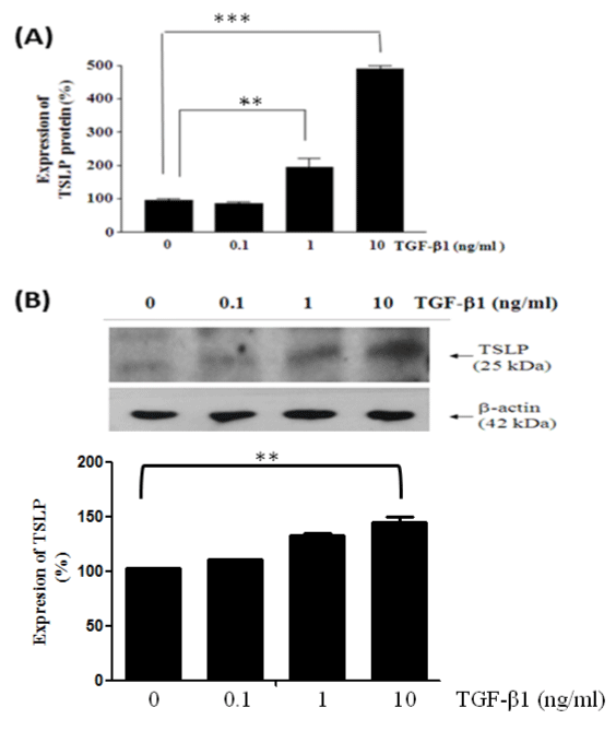
| Figure 1: Effects of TGF-β1 on the expression of TSLP in HFL-1 cells. (A) HFL-1 cells were cultured in serum-free medium (0.5% FBS) with TGF-β 1 (0, 0.1, 1, or 10 ng/ml) for 24 h. Supernatants were collected and subjected to ELISA for detection of TSLP. Experiments were duplicated (n=4) and consistent results were observed. HFL-1 cells were treated with TGF-β1 dose-dependently (0, 0.1, 1, or 10 ng/ml, Figure1B) and lyzed. 100 μg of protein lysate was resolved by 12.5% SDS-PAGE. Western blot was performed by polyclonal anti-TSLP followed by the addition of secondary antibodies (1:4000). β-actin was used as an internal control. Experiments were repeated three times and consistent results were observed. P*<0.05, P**<0.01 versus control (0 ng/ml TGF-β1). |