 |
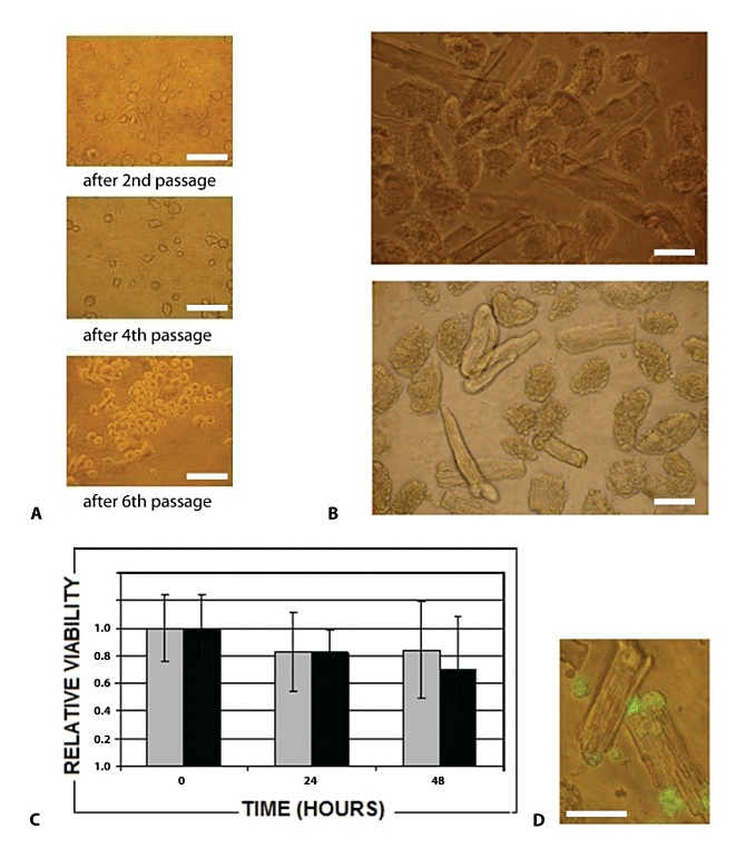
| Figure 1: (A) Morphology of rat BM-MNCs adhered to the plastic bottom on the fifth day after the second, fourth and sixth passages. (B) Morphology of adult myocytes after 24 h in M1 medium (upper image) and in standard IMDM medium with10% FBS (bottom image). (C) Survival rate of adult cardiomyocytes in M1 medium (shadow bar) and standard IMDM medium (black bar). Data are presented as averages ± standard deviation. (D) BM-MNCs (green circular cells) adhere to cardiomyocytes and one of them makes mechanical connection between two cardiomyocytes, Axiovert 40C/CFL Zeiss fluorescence microscope, objective magnification 40×. Scale bars represent 40 μm. |