 |
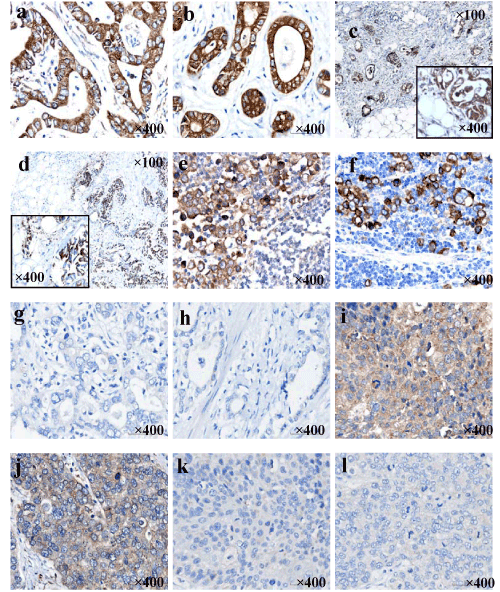
| Figure 2: VEGF and COX-2 IHC staining. (a) strong positive VEGF expression in adenocarcinoma,x400; (b) strong positive COX-2 expression in adenocarcinoma,x400; (c) VEGF positive expression in invasive adenocarcinoma cells in the involved serosa, x100-x400; (d) COX-2 positive expression in adenocarcinoma cells of the serosa,x100-x400; (e) VEGF positive expression in adenocarcinoma cells in the metastatic lymph node,x400; (f) COX-2 positive expression in adenocarcinoma cells in the metastatic lymph node,x400; (g) VEGF negative expression in poorly differentiated adenocarcinoma,x400; (h) COX-2 negative expression in adenocarcinoma,x400; (i) VEGF positive expression in squamous-cell carcinoma,x400; (j) VEGF positive expression in neuroendocrine cancer,x400; (k) COX-2 negative expression in squamous-cell carcinoma,x400; (l) COX-2 negative expression in neuroendocrine cancer,x400. |