 |
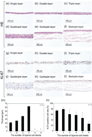
| Figure 1: (a - l) Histological observations of multi-layered cell sheets after 7 days cultivation on culture dishes. (a - f) Photographs of the upper two rows are the hematoxylin and eosin stained cross-sections of the specimens. (g - l) Photographs of the lower two rows are the sections of multi-layered cell sheets stained with anti Ki-67 antibody after 7 days cultivation on culture dishes. Cross-sections were counterstained with hematoxylin for staining nucleus. Photograph (a, g) represent the cross-section of single-layered cell sheet; (b, h), double-layered cell sheets; (c, i), triple-layered cell sheets; (d, j), quadruple-layered cell sheets; (e, k), quintuple-layered cell sheets; (f, l), sextuple-layered cell sheets. (m) The thickness of the specimens appeared on (a) to (f) after 7 days cultivation on culture dishes. The asterisks (*) in the graph show that quintuple- and sextuple-layered cell sheets were unmeasurable state because of their delamination. (n) The average percent of Ki-67 positive cell per 20 times magnified field in multi-layered cell sheets on Day 7. Each data point represents mean ± SD, n = 3. |