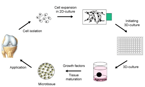
Schematic overview showing the experimental background used in the presented study. The different steps during the generation of scaffold free 3-D cartilage like microtissues are displayed. Cells were isolated from a cartilage biopsy (left) and expanded in monolayer culture (top). Cell aggregation was induced in 96 well plates (right) and microtissues were cultured in agar overlay (bottom).