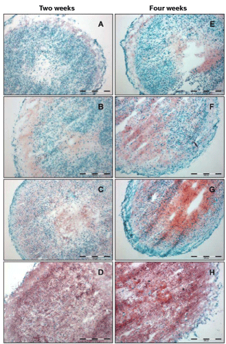
Cryosections of in vitro cartilage-like microtissues stained for the accumulation of proteoglycans with safranin O (red = proteoglycans; cell nuclei = blue) after two and four weeks. A/E: basal medium (bm), B/F: bm + TGF-ß1, C/G: bm + TGF-ß2, D/H: bm + TGF-ß1 + BMP-7. 10x objective; scale bars = 200 μm.