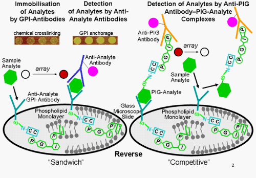
Figure 7: GPI-protein chip for single-parameter analysis.
For the molecular identification of protein analytes in the “Reverse” configuration, they are immobilised by binding to anti-analyte GPI-antibodies/scFvs and then embedded in the phospholipid monolayer coat of the glass microscopic slide. The protein analytes are then detected either by labeled anti-analyte antibodies in the “Sandwich” configuration or by displacement of complexes pre-formed between labeled anti-PIG antibodies and the recombinant PIG-analytes from the anti-analyte GPI-antibodies/scFvs in the “Competitive” configuration. The generation or erasing of the light, fluorescence or luminescence signals, respectively, by the two configurations is read-out from the corresponding microarrays by laser scanners.