 |
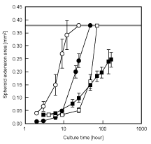
| Figure 4: Change in the cell extension areas of the spheroids transferred onto the collagen-coated dish. A linear threshold shows the maximum spread area under this condition (cells-covered dish). The spheroids of ES cells (○) and 3T3 cells (●) spread readily and covered the dish within 12 and 48 hours after the ST chip was flipped, respectively. The spheroids of HepG2 cells (□) covered the dish within 72 hours. The spheroids of rat primary hepatocytes (■) spread slowly. |