 |
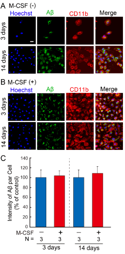
| Figure 4: Phagocytosis of Aβ by adherent cells derived from mouse bone marrow cells. At three and fourteen days after the cultivation of mouse bone marrow cells in the presence (B) or absence (A) of human M-CSF, adherent cells were further treated with Aβ42 for 12 hrs. The adherent cells (nuclei; blue color) expressing CD11b (microglial marker: red color) phagocytosed Aβ (green color). Scale bars = 20 μm. The intensity of Aβ phagocytosed by an adherent cell was measured by image analysis (C). A significant difference of the main effect of treatment × culture day interaction at P = 0.7208 vs. groups cultured in the absence of M-CSF in C. N; number of samples.. |