 |
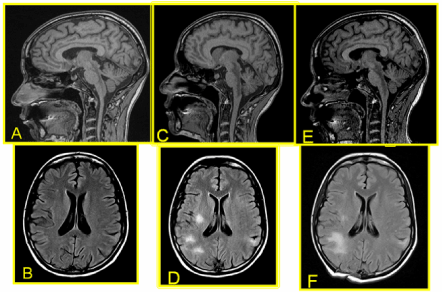
| Figure 1: Before ART: (A) sagittal T1 imaging showing a posterior low signal intensity lesion extended from medulla oblongata to diencephalus; (B) axial Flair without subcortical lesions. Weeks after ART: (C) sagittal T1 imaging with enlargement of the lesion; (D) axial Flair showing high intensity lesions scattered in the subcortical areas. Months after ART:(E) sagittal T1 and (F) axial Flair images with mild improvement. |