 |
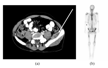
| Figure 1: a) Transverse section abdominal CT showing left iliacus abscess (white arrow) four days after presentation described in the main text. There was no bowel wall thickening or anatomical abnormality apparent in this scan. b) Delayed image 99Technetium bone scan anterior view two weeks after commencing intravenous antibiotics. There is increased isotope uptake in both iliac bones, proximal right humerus and both femurs. These changes are consistent with osteomyelitis. |