 |
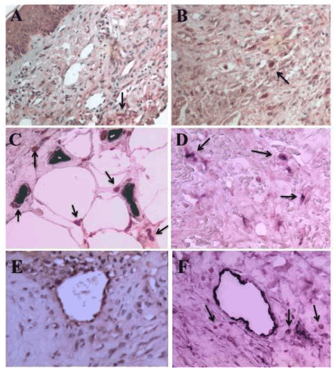
| Figure 1: Detection of BP1 and FGF-2 in Kaposi’s sarcoma skin lesions by immunohistochemistry. Panels A and B show localization of BP1 protein (red color) in AIDS-KS skin lesions pointed by the black arrows (A, 200X; B, 500X). Panel C shows capillary endothelial cells stained positive with vWF (dark color) located in close proximity to infiltrating cells expressing BP1 (red color) and pointed by the black arrows (500X). Panel D shows co-localization of BP1 (red color) in infiltrating mononuclear cells stained also with an antibody against the CD-68 antigen (dark color) in a KS skin lesions (500X). Dual BP1 and CD68 positive cells are pointed by the black arrows. Panel E, shows the localization of FGF-2 (red color) in an AIDS-KS skin lesion. FGF-2 was localized in endothelial cells, infiltrating spindle cells, and the extracellular matrix (500X). Panel F shows vWF positive endothelial cells in an AIDS-KS skin tumor (dark color), and infiltrating BP1 positive cells (red color) surrounding the vessels. Black arrows point to the BP1 positive cells (500X). |