 |
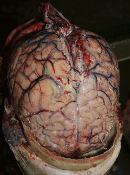
| Figure 3a: The photograph shows the gross appearance of the brain within the cranial cavity following sawing of the skull cap. There is a scanty purulent exudate seen in the subarachnoid space (black arrow). The surface vessels are congested and the brain is visibly swollen showing signs of increased intracranial pressure. |