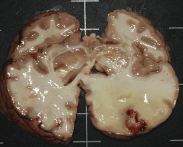
The histopathological examination of the brain showed mixed aetiology meningoencephalitis (acute bacterial infection and Cryptococcus yeast forms). A focal area in the parenchymal corresponding with a cryptococcoma showed surrounding gliosis and hemorrhage. The yeast forms stained positive with PAS stain. The ZN stain was negative.