 |
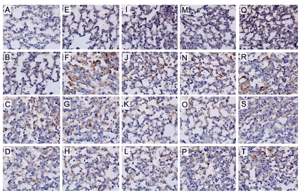
| Figure 2: Photomicrographs of the results of immunohistochemical analyses of lung tissue to detect IL-2 (panels A–D, 400X), IL-4 (panels E-H, 400X), IL-13 (panels I–L; 400X) IFN-γ (panels M–P, 400X) and iNOS (panels Q-T, 400x) positive cells from guinea pigs exposed only to saline (panels A, E, I , M and Q) or ovalbumin (panels B, F, J, N and R). The guinea pigs from OT1 (panels C, G, K, O and S) and OT2 (panels D, H, L, P and T) groups are also represented. |