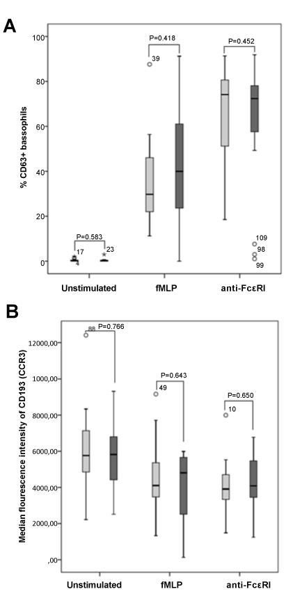
Figure 3: Percentages of CD63 positive activated peripheral blood basophils
(Panel A) and median fluorescence intensity of CD193 (CCR3) expression
(Panel B) on activated peripheral blood basophils, in patients with mastocytosis
(n=19, dark grey boxes) and in healthy individuals (blood donors) (n=19, light
grey boxes), at the baseline (unstimulated) and after stimulation with fMLP or
with anti-FcεRI.
Boxes extend from the 25th to the 75th percentiles; the line in the middle and
the vertical lines represent median values and 95% confidence intervals,
respectively. No significant differences (P>0.05) were observed between the %
of CD63+ basophils and the MFI of CD193 (CCR3) expression on basophils in
patients and in controls using the Mann-Whitney U-test. |
