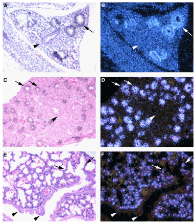
In situ hybridization was used to identify sites of SP-C expression in the developing mouse lung: embryonic day (ED) ED13 (panels A, B), ED15 (panelsC, D), ED18 (panelsE, F). A, C, E are bright field images to visualize tissue morphology. B, D, F are corresponding dark field image where hybridization of the SP-C riboprobeto SP-C mRNA appears as light granules. Arrowheads indicate central airway epithelia, arrows indicate peripheral extension of the growing airway(s)epithelia where SP-C is expression is sustained. Weak SP-C expression is detected in cells of the primitive airways of the ED13 lung rudiment where branch formation has begun. SP-C expression is already decreased in the central most tubule (arrowhead panels A, B). The relative expression SP-C has increased and localizes to the extendingepithelium in ED15 lung. As branching morphogenesis proceeds SP-C expression is silenced in cells lining thelarger central airways(arrowhead) and restricted to the most peripheral aspects of the invading epithelium. By ED18 lung mesenchyme has thinned and septation has begun. SP-C expression is extinguished in the conducting airway epithelia and is only detected as strong focal sites of expression in the alveolarizing compartment.All images 20X original magnification.