 |
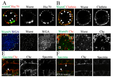
| Figure 4: Wurst is involved in clathrin-mediated endocytosis (A-D) Immunofluorescent analysis using anti-clathrin, anti-Chc, anti-Hsc70, anti-WurstC (recognizing the C-terminus [13]), and anti-WurstN (recognizing the N-terminus, see materials and methods) antibodies in Drosophila SL2 cells (A,B) and in membrane sheets of SL2 cells (C,D). The anti-clathrin antibody recognizes Chc and Clathrin light chain. (A,B) Wurst is localized in a vesicular like pattern in SL2 cells, which partially co-localize (arrowheads) with Hsc70 (A) and clathrin (B). (C) Wurst appears distributed in small clusters in the membrane sheet of a SL2 cell. Wheat germ agglutinin (WGA) selectively recognizes sugar residues at the plasma membrane surface. White dashes mark the membrane sheet. (D) Magnification of the membrane sheet reveals Wurst (green) and Chc (red) overlap (rectangles). (E,F) Confocal images using anti-Chc and anti-α-Spectrin antibodies in Drosophila stage 17 embryos. The α-Spectrin marks cell membranes. In wild-type tracheal cells Chc is found at the membrane and in addition in the cytoplasmic cell cortex (arrowhead in E). In contrast, in btlG4 driven UAS-RNAi-wurst tracheal specific knock-down embryos, Chc is accumulated at the apical plasma membrane of tracheal cells (arrowheads in F). Scale bars indicate 5μm.. |