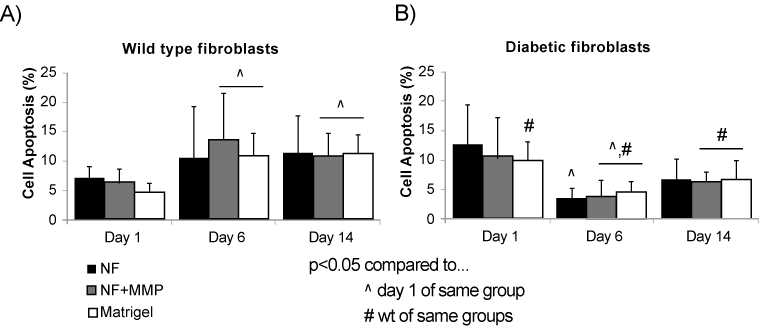
a) The percentage of apoptotic wild type (wt) fibroblasts was less than 15% for in NF, NF+MMP and Matrigel scaffolds at days 1, 6 and 14. While no difference was observed in apoptosis in NF scaffold, there was a significant increase in apoptosis in both NF+MMP and Matrigel from day 1 to days 6 and 14.
b) The percentage of apoptotic diabetic (db) fibroblasts was less than 15% for in NF, NF+MMP and Matrigel scaffolds at days 1, 6 and 14. Initial (day 1) apoptosis levels in db fibroblasts were significantly higher and decreased at day 6 in all scaffolds. ^ p<0.05 when compared to day 1 samples of same experimental group, # p<0.05 when compared to wt samples of same experimental groups.