 |
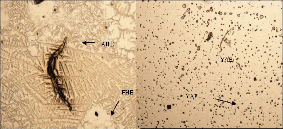
| Figure 3: Phase contrast and differential interference contrast (DIC) photomicrographs of live hemocytes of F.indicus (A, B). Phase contrast images of hemocytes showing agglutination with HB RBC. Note that the Agglutinated erythrocytes & haemocytes (AHE) appeared darker and markedly lost their surrounding bright rings as compared to free (FHE) or attached (YAE) erythrocytes. DIC images of the same area. |