 |
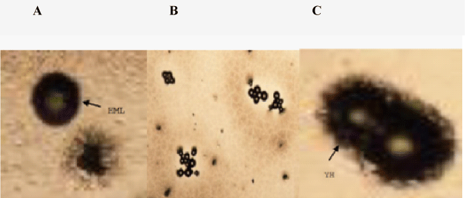
| Figure 4: Rounded haemocytes were incubated with yeast cells and the attachment with few yeast cells to haemocyte surface, free yeast cells were found in monolayer (Haemocyte monolyer HML) (A). F. indicus haemocyte attached with 5 yeast cells intracellularly that were surrounded by bright rings and visible under phase optics (B). Ingested yeast cells emerge darker and lost their surrounding bright rings obviously as compared with free or extracellularly attached yeast cells (yeast haemocytes –YH) (C). |