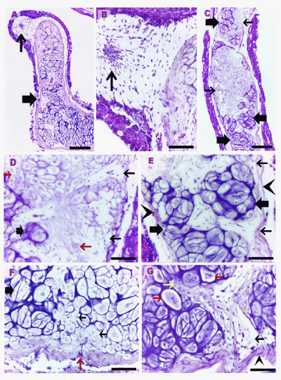
Paraffin sections of air respiratory organ of catfish measured 13.4 (A), 13.2 (C-E), 14.1 (F), 14.9 (G) cm in length, stained with H&E. A, B: The thin arrows refer to the areas of mesenchymal cell condensation. C: The thin arrow point to the differentiating chondrocytes; Note little extracellular cartilage matrix around the differentiating chondrocytes. The thick arrows refer to the cartilaginous masses. D, E, G: The thin arrows refer to areas of the cellular invasion to the growing cartilage. The red arrows refer to differentiating chondrocytes in "D", chondrocytes doublets in "G" and the invading perichondrial mesenchymal cells to the growing cartilage in "F". The yellow arrow refers to the differentiating chondrocytes. The arrow heads refer to the perichondrium. Scale bars represent 80 μm in "A, C", 50 μm in "B, D-G".