 |
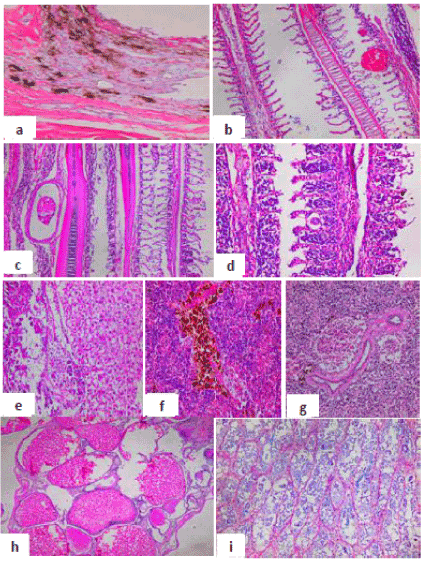
| Figure 4: Histopathological changes in Oreochromis niloticus collected from site II showed: a: Skin with severe hyperplasia and hypertrophy in the epidermal layer and excessive accumulation of melanophores in the dermal layer. b: Gills showed edema with epithelial lifting, telangiectasia. c&d: Gills showed lamellar fusion, epithelial hyperplasia, proliferation of hypertrophic cells, external parasites in between the gill tissues surrounded with fibrous connective tissue sheet. e: Liver showed severe degenerative and necrotic changes in the hepatocytes and in the pancreatic tissues with aggregation of mononuclear inflammatory cells. f: Spleen showed congestion and hyperactivation of melanomacrophes cells. g: Spleen showed depletion of the lymphocytic tissues, congestion and hyperplasia in the wall of blood vessels. h: Ovary showed in ripe stage with liquefaction of cytoplasm of oocyte, nucleus loses and degeneration in wall of oocyte with liquefaction of the yolk sphere with large vacuoles of ripe stage and irregular wall of oocytes. i: Testis showing degeneration and necrosis in the primary spermatocytes in semniferous tubules and decreased number of spermatogenic cells. (H&E,X40). |