 |
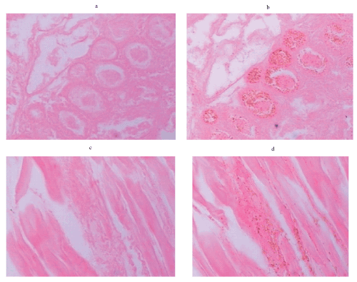
| Figure 3: Representative immunohistochemical detection of V. harveyi infected tissues from dead P. monodon following exposure to V. harveyi 639. The (a,b) hepatopancreas and (c,d) heart muscle after staining by haematoxylin and eosin(H & E) while the right panels (b,d) in each case were also stained with indirect immunoperoxidase using the VH3-3H monoclonal antibody as a probe. Thus, brown dots show the presence of V. harveyi 693. Images shown are representative from three independent shrimp and examined under Light microscope (x 1,000 magnification). |