 |
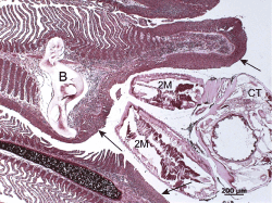
| Figure 1: Light micrograph of the attachment of the lernaeopodid copepod Salmincola californiensis to a gill lamella of a spawning pink salmon (Oncorhynchus gorbuscha). The lamella is distended by the anchoring bulla (B) and epi- thelial hyperplasia (arrow) is evident adjacent to the site of attachment. Parasite structures also visible are the second maxillae (2M) and cephalothorax (CT). Gram stain. |