 |
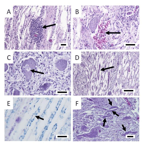
| Figure 2: Histopathological sections of the central nervous system of affected yellowtail. A-D and F: H & E stain, E: Kluver-Barrera stain. Arrows show the glial nodule (A), blood congestion and hemorrhages (B), nerve cell necrosis with neurophagia (C), swollen eosinophilic nerve fibers (D), demyelination caused by degenerative neurons (E), and clusters of elongated myxosporean plasmodia (F) in the brain and spinal cord. Scale bars = 30 μm |