 |
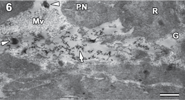
| Figure 6: Severe distortion in the cytoplasmic membrane was seen at higher concentration of 110μgL-1 characterized by electron dense cytoplasm and swollen Nuclei (N) with indistinct Nucleolus (NU), vacuolation of nucleus (arrow) and disintegration of cytoplasmic inclusions (arrowhead). Golgi body (G), Lysosomes (Ly), microvilli (Mv), lipid droplet (L), scale bar 0.5μm. |