 |
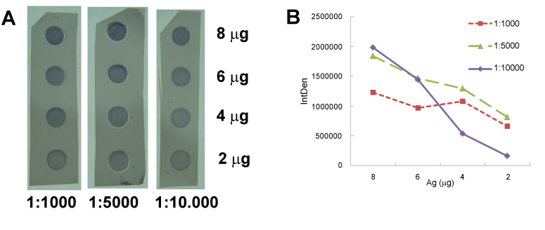
| Figure 2: (A) Dot-ELISA affinity test on PVDF membrane: Optimization from various amounts of antigen (Amyloid β), 2-8 μg, and concentrations of secondary antibody, 1:1000 or 1:5000 and/or 1:10000, DAB staining. (B) Densitometric analysis of spots on blotting membrane: Plot of amount of antigen vs. integrated density of spots. |