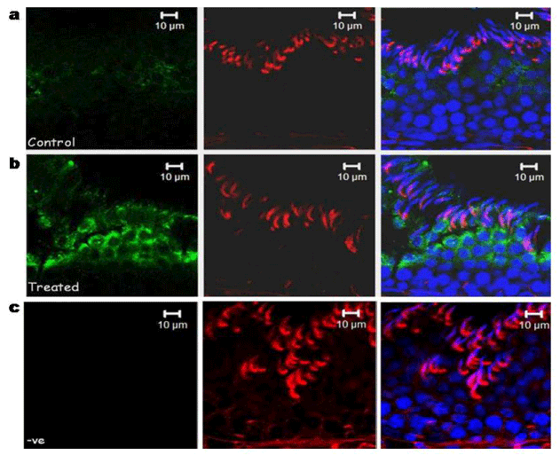
Panel a shows localization in the control group (normal spermiation) and panel b shows localization in the treated group while subpanel c show negative control with rabbit IGg instead of primary antibody.
Green fluorescence=primary antibody (calnexin), red fluorescence=phalloidin (F-actin marker), blue fluorescence=DAPI (nuclear stain), Bar=10 μm.