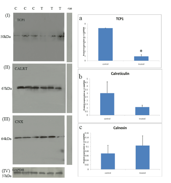
Panel I, II, III represents western blotting of the TCP-1, calnexin and calreticulin proteins respectively from tubules at stages VII-VIII in control group and treated group. –ve denotes negative control without primary antibody.
Subpanel a,b,c denotes graphical representation of expression of TCP-1, calnexin and calreticulin proteins respectively in control (normal spermiation) and treated (failed spermiation) groups. Data n=3 animals, *=significant (p ≥ 0.02).