 |
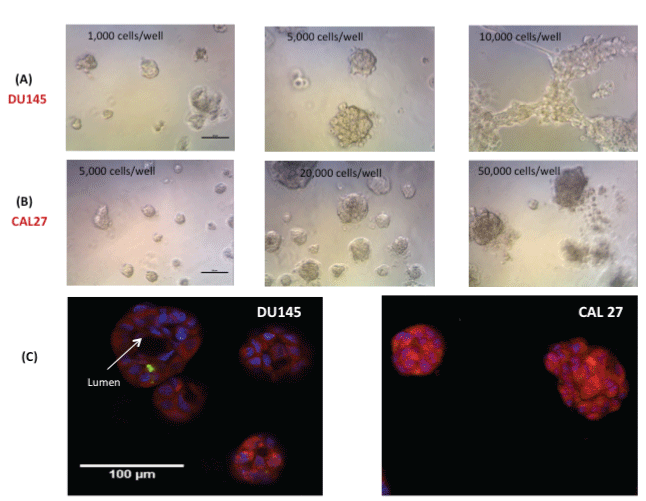
| Figure 1: Optical microscopic images of 3D spheroids on Matrigel of DU145 and CAL27 cells. (A) DU145 cells at initial cell number ranging from 1,000 to 10,000 cells/well, (B) CAL27 cells at initial cell number ranging from 5,000 to 50,000 cells/well, at day 4; And (C) the fluorescent confocal microscopic images of the structure of DU145 spheroids and CAL27 spheroids on Matrigel. The spheroids were stained with10 µg/ml Hoechst dye, 10 µM cell tracker red, 1:1000 Cell Tox Green in 100 µl of fresh base medium at 37°C for 30 min. Scale bar in (A), (B), and (C)=100 mm |