|
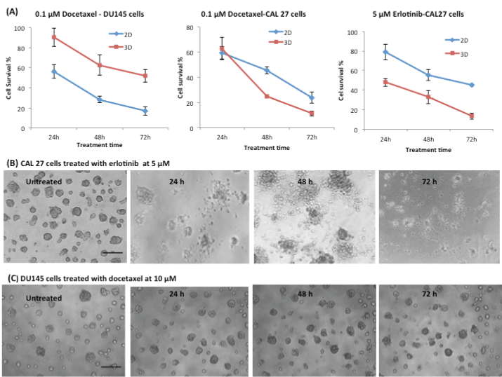
| Figure 4: Cellular responses of 3D and 2D cultured DU145 and CAL27 cells. (A) The differences in cell survival between 3D and 2D cultured cells of DU145 upon treatment of 0.1 µM docetaxel, and of CAL27 upon treatment of 0.1 µM docetaxel and 5 µM erlotinib, for 24, 48, and 72 h, measured by MTT assay. (B) Optical microscopic images of CAL27 spheroids upon treatment of 5 µM erlotinib for 24, 48, and 72 h, along with the untreated one. (C) Optical microscopic images of DU145 spheroids upon treatment of 10 µM docetaxel for 24, 28, and 72 h, along with the untreated one. Scale bar in (B) and (C)=100 µm. |