 |
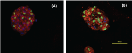
| Figure 5: Fluorescent confocal microscopic images 3D spheroids of CAL27 and DU145 cells. (A) CAL27 spheroids treated with 0.5 µM erlotinib for 72 h, and (B) DU145 spheroids treated with 41.1 µM rapamycin for 48 h. The spheroids were stained with10 µg/ml Hoechst dye (nucleus of live cells), 10 µM cell tracker red (cytoplasm), 1:1000 Cell Tox Green (nucleus of dead cells) in 100 µl of fresh base medium at 37°C for 30 min. Cells were stained for cytoplasm (red), nucleus of live (blue) and dead (green) cells. |