 |
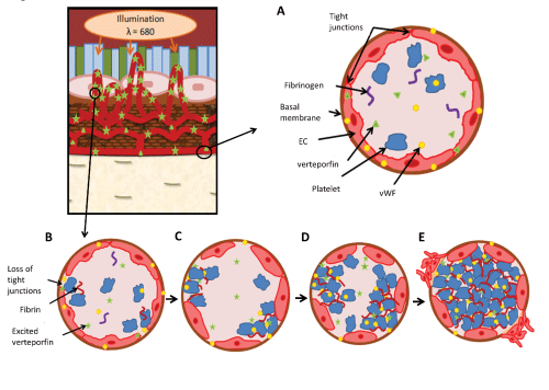
| Figure 1: An overview of the vascular effects of photodynamic therapy (PDT) leading to vasoconstriction, blood flow stasis and thrombus formation. (A) Magnification of a normal choroidal blood vessel and (B-E) magnifications of a choroidal neovessels being illuminated. (A) Choroidal blood vessel where verteporfin remains in the inactive form due to lack of illumination and no effects of PDT can be seen. Under normal circumstances, the endothelial cells (EC) line blood vessels with a close association to the basement membrane, and are connected to each other through tight junctions. (B) Light exposure in the choroidal neovessels activates verteporfin initiating EC stress and various cellular responses, including the retraction of ECs resulting in the loss of the tight junctions between cells and the separation of cells from the basement membrane. (C) The interaction of blood with collagen and other factors in the basement membrane results in the activation of the clotting cascade, including the activation of platelets and the release of von Willebrand factor (vWF). (D) Cellular responses result in vascular constriction and the stabilization of the blood clot by fibrin, which is produced from fibrinogen as a part of the clotting cascade. (E) Vessel occlusion due to vaso-constriction and clot formation results in the activation of the angiogenic switch, results in the activation of migration and the sprout formation of ECs. |