 |
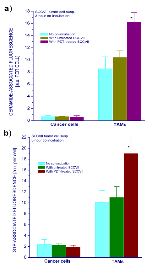
| Figure 3: Ceramide and S1P levels in cancer cell and TAM populations from SCCVII tumors following co-incubation with PDT-treated SCCVII cells separated in culture inserts. Cells from untreated SCCVII tumors were plated in 35 mm Petri dishes and co-incubated 3 hours with culture inserts containing in vitro grown SCCVII cells that were either untreated or PDT-treated (as described for Figure 1). After the co-incubation, cells were collected and stained for identifying TAMs (as described for Figure 2), while anti-ceramide (a) or anti-S1P antibodies (b) were used for assessing cellular levels of these sphingolipids based on flow cytometry. The bars denote SD; *=statistically significant difference from the values with the same type of cells not co-incubated with SCCVII cells. |