 |
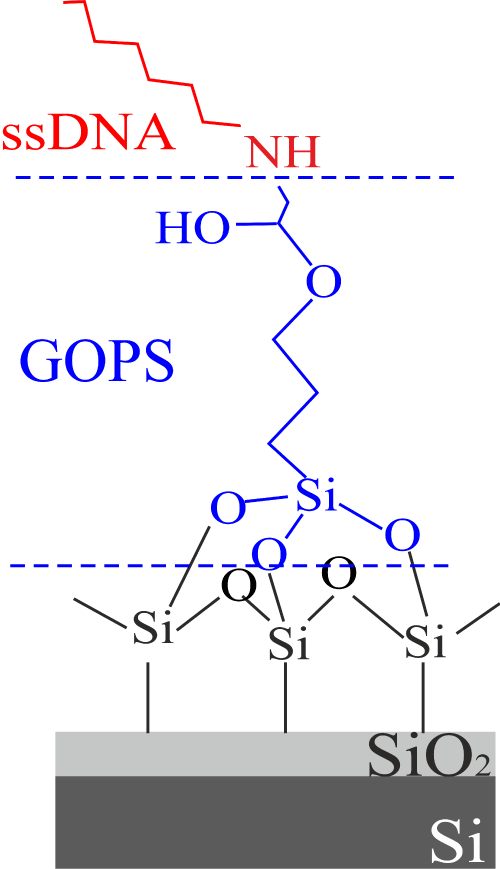
| Figure 5: Schematic structure of the GOPS immobilization protocol. The GOPS structure is in blue separated by horizontal dashed lines from the activated oxide (below the GOPS) and the immobilized biological molecule (in red above the GOPS, amino-terminated ssDNA is shown as an example). |